Title : THE CYSTOHEPATIC MESENTERY : A NEW ANATOMICAL LAPAROSCOPIC SURGICAL PERSPECTIVE
By : Abdelghany Elshamy
Professor of Surgery
Consultant Of Pancreatobiliary, Laparoscopic and Endoscopic Surgery. Ain Shams University. CAIRO. EGYPT.
THE CYSTOHEPATIC MESENTERY .
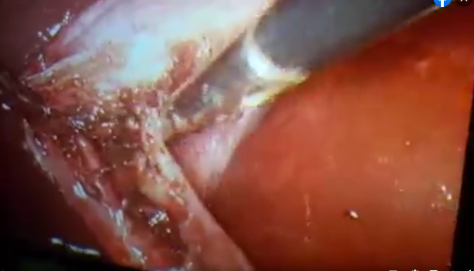
This is our Current personal laparoscopic anatomical perspective of the CYSTOHEPATIC TRIANGLE or TRIANGLE OF CALOT .
During laparoscopic cholecystectomy dissection of this triangle or i think is better to be termed The CYSTOHEPATIC MESENTERY (CHM) or CYSTOHEPATIC MESENTERIC SPACE (CHMS), we have observed that this small mesentery is formed of an anterior visceral peitoneal layer , then the fatty layer in the middle , then the posterior peritoneal layer . So , it is formed of three layers (trilaminar), in which the fatty layer is invested between the anterior and posterior visceral peitoneal layers. The anterior peritoneal layers is reflected just around the cystic duct or a couple of millimeters below it to become the posterior layer , so to visualize the cystic duct we have to dissect mainly above it and we continue dissection around it pealing the fat for proper visualization. The contents of this space include the cystic artery or better to refer to it as cystic blood vessels (as there are frequently anterior and posterior branches or sometimes multiple small vessels) , the cystic lymph node , and other minute lymphatics and nerve fibres.
Laparoscopic or Endoscopic implications of this mesenteric triangle or mesenteric space :
During laparoscopic cholecystectomy we usually start dissection in this space anteriorly by dissection of the anterior visceral peitoneal layer to visualize the cystic duct, and cystic vessels. Then the middle fatty layer is visualized and dissected , then the posterior layer is dissected . I prefer to dissect the posterior layer after reflecting the neck of the gallbladder or the Hartmann’s pouch anteriorly and medially and dissect it better from posterior under vision or it can be dissected from anterior after dissection of the fatty layer.
Most laparoscopic surgeons have observed that the cystic lymph node of Lund is considered as a landmark of the cystic artery or its anterior branch.
During dissection of the posterior layer of CYSTOHEPATIC MESENTERY we have to be causious to avoid bleeding from a posterior branch of cystic artery or bleeding from right hepatic artery with a caterpillar course or accessory right hepatic artery . Usually we complement the anterior dissection with dissection of the visceral peritoneal layer from posterior to visualize anomalous course or accessory right hepatic or large posterior cystic vessel to avoid bleeding.
The cystic lymph node may be small and sometimes large according to the inflammation, it may be partially dissected and retracted inferomedially towards the common hepatic duct just to visualize the cystic artery to deal with it near to the gallbladder and away from the common hepatic duct as this is more safe avoiding injury to the bile duct or bleeding in this space , this lymph node may somtimes be removed to visualize the cystic artery, but I prefer to preserve it as possible .
Impacted stones or Mirizzi syndrome will lead to deformed or partial or near total obliteration of Calot triangle or CYSTOHEPATIC TRIANGLE and that is why it is better to be termed CYSTOHEPATIC MESENTERY or CYSTOHEPATIC MESENTERIC SPACE or CYSTOHEPATIC SPACE and the Critical view of Safety is difficult to be achieved.
Also , acute and chronic inflammation will lead to inflammation in the CYSTOHEPATIC MESENTERY and it Will be shortened and thickened and deformed making the space smaller or even about to be obliterated with difficulty in dissection and identification of structures due to fibrosis, excessive vascularity and thickening and shotening of Cystohepatic Mesentery.
The dissection should be away from the Common hepatic duct and the common bile duct should not be dissected except in cases of CBD exploration or for proper visualization before ligation or clipping of cystic duct.
Value of this detailed laparoscopic or microscopic Anatomy :
Our Aim is Safe Cholecystectomy in the era of laparoscopic surgery. This can be achieved with current understanding of detailed laparoscopic anatomy of the Cystohepatic triangle OR Cystohepatic Mesentery.
This will facilitate identification of the structures with great precision avoiding injury of common hepatic duct, Common bile duct and, anomalous right hepatic artery and bleeding with its consequences in this space and important area during laparoscopic cholecystectomy .
These anatomical consideration can be applied to open cholecystectomy as well.
SUMMARY AND CONCLUSION :
In the era of Laparoscopic Surgery ,
The detailed Laparoscopic or Endoscopic anatomy of the Calot’s triangle or personally I prefer to term it CYSTOHEPATIC MESENTERY (CHM) or CYSTOHEPATIC MESENTERIC SPACE (CHMS) is the basis for successful standardized laparoscopic cholecystectomy in the era of minimally invasive surgery.